
 PollyMap
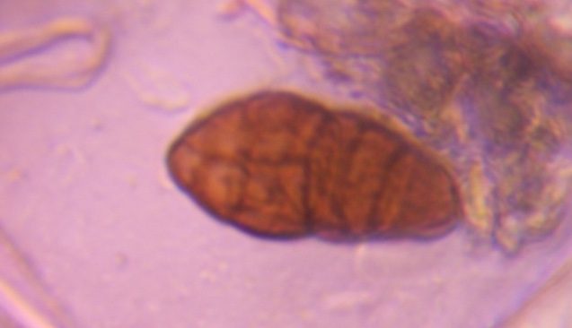
Ulocladium mold spore photographed through the microscope in the PollyKit lab — verified in Arizona.
More verified photos of Ulocladium
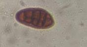
About ulocladium mold spores
Egg-shaped dark spores closely related to Alternaria, strongly associated with water-damaged materials.
Where it grows
- Wet drywall, wallpaper and paper products
- Damp textiles and carpet backing
- Soil, dung and decaying plants
- Outdoor plant debris
How to recognize it
Dark brown, broadly egg-shaped to obovoid spore with both crosswise and lengthwise internal walls and a smooth to faintly roughened surface. No beak, which is the quickest separation from Alternaria.
Typical published size: 15–30 µm
Commonly confused with
- Alternaria — tapering beak at one end
- Stemphylium — pronounced central constriction
Allergy relevance
Moderately allergenic and, because it needs sustained wetness, a useful indoor water-intrusion marker as well as an allergen.
Where it's found
PollyMap has verified specimens of Ulocladium from Arizona.
Worldwide; indoors wherever materials stay wet.
Also searched as: ulocladium spore, ulocladium under microscope, water damage mold spore.
Related mold specimens
Citing this specimen: PollyMap Specimen Library. “Ulocladium — verified microscope specimen.” pollymap.com/library/specimen/ulocladium-mold-spore-62dada
Expert discussion
No published comments yet. If you study palynology or mycology, we'd value your read on this specimen.