
 PollyMap
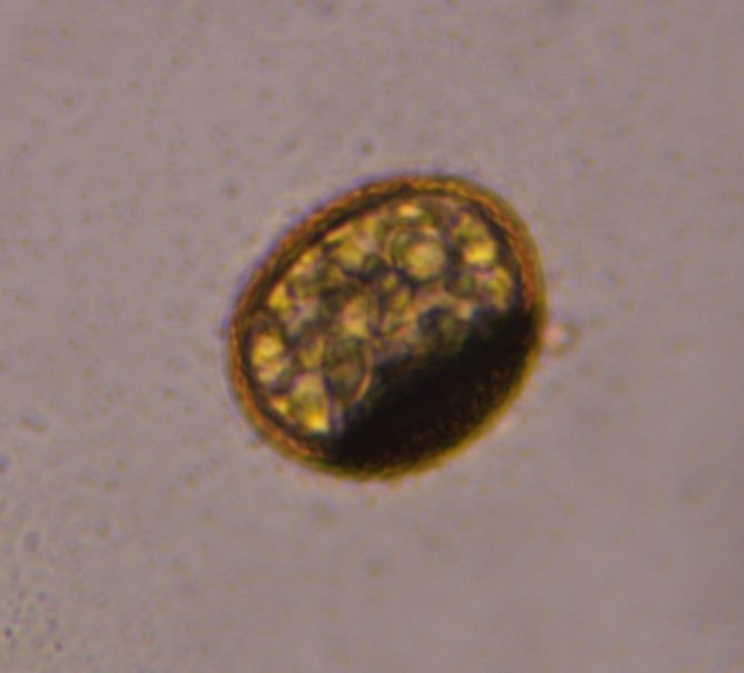
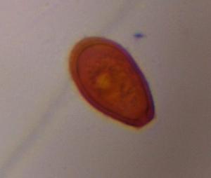
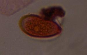
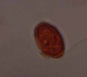
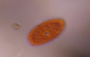
Rust spore mold spore photographed through the microscope in the PollyKit lab.
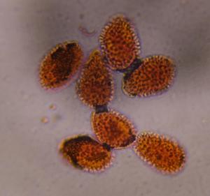
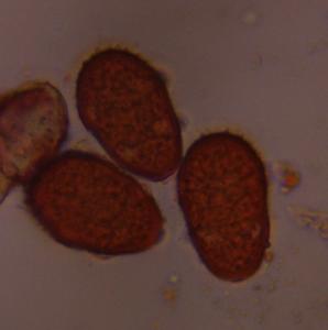
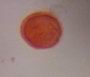
More verified photos of Rust spore


About rust spores
Spores of plant rust fungi, shed in clouds from infected leaves and stems.
Where it grows
- Rust-infected grasses and cereals
- Garden and ornamental plants with rust
- Native shrubs and trees
How to recognize it
Orange-brown to dark brown, thick-walled spore that is often warty or spiny, sometimes with a stalk remnant; urediniospores are round and echinate while teliospores are darker and heavier walled.
Typical published size: 15–35 µm
Commonly confused with
- Smut spores — usually smaller and more uniformly granular
- Epicoccum — internal walls
Allergy relevance
Considered a mild to moderate allergen, mostly relevant to people with heavy outdoor or agricultural exposure.
Where it's found
Wherever host plants grow, across the United States.
Also searched as: rust spore, rust teliospore, urediniospore, plant rust under microscope.
Related mold specimens
Citing this specimen: PollyMap Specimen Library. “Rust spore — verified microscope specimen.” pollymap.com/library/specimen/rust-spore-mold-spore-c3bf36
Expert discussion
No published comments yet. If you study palynology or mycology, we'd value your read on this specimen.